Instruments
MASCOT carries 4 scientific instruments:
![]()
MASCOT's MicrOmega, Cam, Mara, and Mag insutrments - Credits DLR
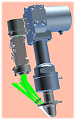 MicrOmega, a hyperspectral infrared microscope for in situ mineralogical analyses of the ground, developed by the IAS (Institut d'Astrophysique Spatiale)
 MASCAM, a multispectral wide field camera to provide geological images of the visited sites, developed by DLR (Berlin) (German Space Agency)
 MasMag, a magnetometer, developed by Braunschweig Technology University
 MARA, a radiometer to determine the surface temperature and the thermal inertia of the asteroid, developed by DLR (Berlin) (German Space Agency)
MicrOmega
The MicrOmega instrument on Hayabusa2 works the same as the MicrOmega instrument developed for Phobos Grunt, itself a prototype of the instrument being developed for ExoMars. It is summed up in the figure below:
![]()
MicrOmega functional principle
A sample directly on the ground is illuminated with a monochromatic light by an illumination system, composed of a lamp (white light) and a monochromator, based on an AOTF (acousto-optical tunable filter): for one excitation frequency of the electrodes mounted on the AOTF crystal, producing an acoustic wave at a given wavelength, the radiation coming out of the crystal is monochromatic, at a wavelength directly function of the one of the acoustic wave. A two-dimensional image is then acquired by the detection system (composed in HgCdTe), cooled by a miniaturized cryogenic machine. A second wavelength is then chosen, by changing the excitation frequency, and a second image is acquired.
Thus, step by step, by successive wavelength stages obtained by simple scanning frequency of the signal applied to the electrodes of the crystal, a spectral scanning is carried out: a three-dimensional image-cube (x,y,![]() ) is acquired for this sample, where each pixel of the image has a spectrum with as many spectral points as the number of stages in the frequency scanning.
) is acquired for this sample, where each pixel of the image has a spectrum with as many spectral points as the number of stages in the frequency scanning.
![]()
Three-dimensional image-cube (x,y,![]() )
)
The scanned frequency area ranges from 0.95 to 3.65 µm, in 365 spectral channels: in this area, and with this resolution, the spectral signatures of most desired mineralogical and molecular components have identifiable diagnostic signatures.
![]()
Image and spectra of a sample of pyroxene, kaolinite and nontronite, performed with the MicrOmega prototype:
pyroxene (1, blue); nontronite (2, green); kaolinite (3, red)
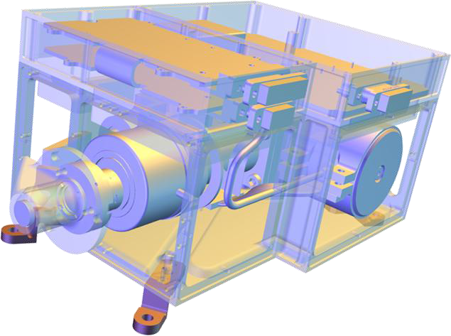
With a total mass of 2 kg, MicrOmega is made up of the following sub-systems:
- a detection sub-system and its cryogenic machine (Sofradir)
- an illumination sub-system made up of
- a lamp
- a RF generator (Erems)
- an AOTF (Gooch&Housego)
- an electronics sub-system (Steel) made up of
- a proximity electronics to control the detector, the cryogenic machine, the lamp and the thermal sensors
- a deported electronics in the Ebox
- an optics sub-system made up of